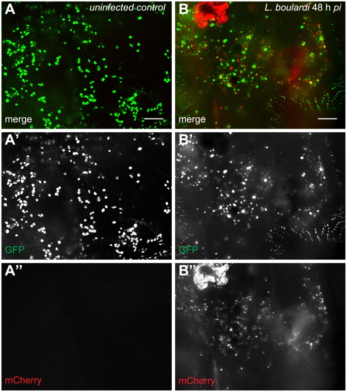
Fig 3. Activated plasmatocytes.
Sessile hemocytes of (A-A”) uninfected Me/w control larvae and (B-B”) L. boulardi-infected Me/w larvae 48 h after infection. The images show the terminal segments of two Drosophila larvae. In uninfected controls, plasmatocytes were the predominant sessile cell type, whereas in infected animals activated plasmatocytes were prevalent. The merge and the independent channels are shown separately. Scale bars 50 μm.

