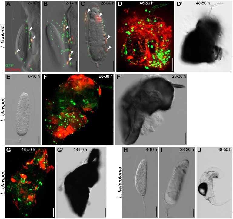
Fig 5. Chronological events during the cellular immune response on the surface of wasp eggs.
Me/w heterozygous Drosophila larvae were infected by (A-D’) L. boulardi, (E-G’) L. clavipes or (H-J) L. heterotoma, and wasp eggs and wasp larvae were dissected and imaged at the indicated time points. L. boulardi eggs were always attached to the gut. Already 8–10 h after infection, plasmatocytes were visible on the wasp eggs and some started to express mCherry, transforming into activated plasmatocytes. The number of plasmatocytes and activated plasmatocytes on the wasp egg increased during the course of infection. In most cases, L. boulardi wasp larvae hatched around 32 h after infection, and some of them were later killed by the cellular immune system. No hemocytes attached to L. clavipes eggs early after infection. Eggs were encapsulated and melanized 28–30 h after infection, and wasp larvae rarely hatched from these eggs. No hemocytes ever attach to the freely floating eggs of L. heterotoma, and the wasp larvae of this species hatched around 38 h after infection.

