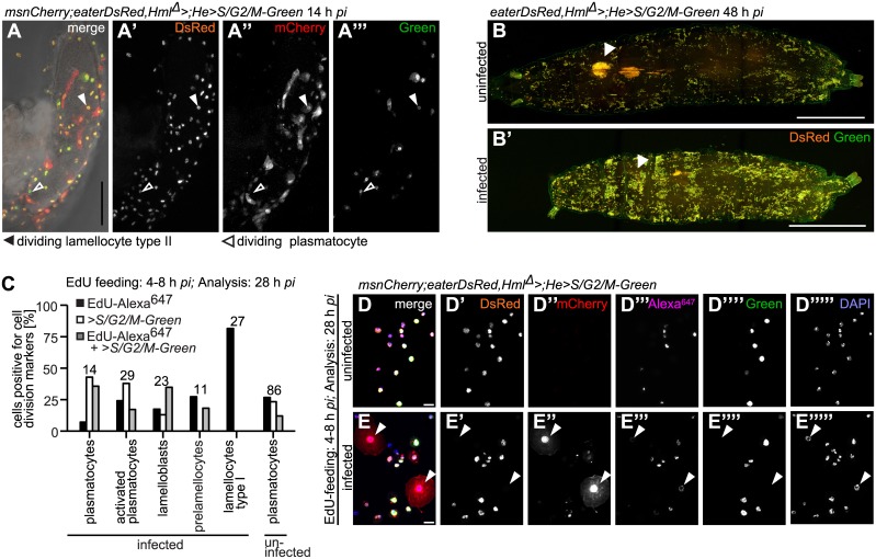
Fig 7. All larval hemocyte types are able to divide, except for lamellocytes.
(A-A”‘) Hemocytes were able to divide on wasp eggs. Arrowheads point to an example of a dividing activated plasmatocyte and a dividing plasmatocyte. Scale bars 50 μm. (B-B’) Sessile blood cells divided in control and L. boulardi-infected larvae 48 h after infection. eaterDsRed;Hml Δ > was crossed with w;He>S/G2/M-Green to obtain eaterDsRed;Hml Δ >;He>S/G2/M-Green. Observe that due to the genotype only plasmatocytes are visible after infection! The number of sessile cells increased after a wasp infection. In the wasp-infected larva, the primary lymph gland lobes were no longer present, whereas they were still visible in the control larva. Arrowheads point to the locations of the primary lymph gland lobes. Scale bars 100 μm. (C) Quantification of dividing cell types after EdU feeding 4–8 h and analysis 28 h after infection of uninfected and L. boulardi-infected larvae. Hemocytes of offspring of crosses of msnCherry;eaterDsRed,Hml Δ > to w;He>S/G2/M-Green were imaged and analysed for the incorporation of EdU-Alexa647, and/or the expression of S/G2/M-Green. The total numbers of evaluated cells per cell type are indicated above the bar plots of each hemocyte type (pc: plasmatocytes). All cell types incorporated EdU. All cell types but lamellocytes expressed Green. All cell types except for lamellocytes incorporated EdU and simultaneously expressed >S/G2/M-Green. The gating strategy is explained in S9 Fig. (D-E”“‘) Representative images of cells analyzed in (C). Arrowheads point to lamellocytes. None of the lamellocytes expressed Green (E”“), but one lamellocyte that had incorporated EdU is shown (E”‘). Scale bars 50 μm.

