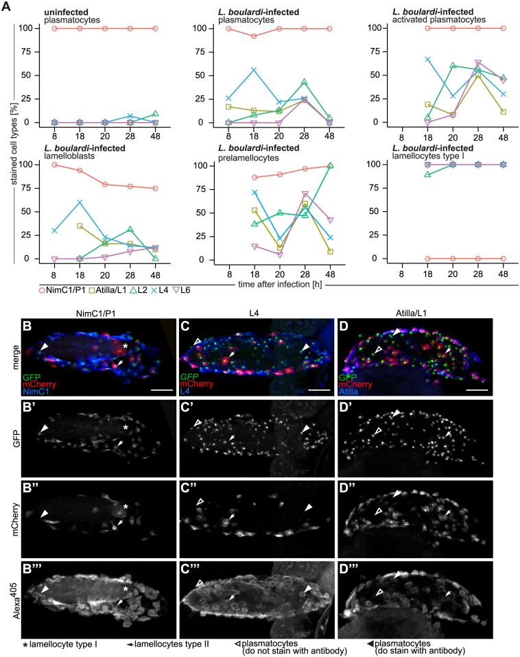
Fig 10. Antibody staining of hemocytes fixed on glass slides and on L. boulardi eggs.
(A) Staining of Me-expressing hemocytes of age-matched control and L. boulardi-infected larvae with NimC1/P1, Atilla/L1, L2, Myospheroid/L4, and L6 antibodies at 8, 18, 20, 28, and 48h after infection. Individual hemocyte types are shown in separate graphs. Hemocytes of uninfected control larvae were primarily plasmatocytes and stained mainly with NimC1/P1 antibody at all time points. All blood cells of infected larvae stained with NimC1/P1 as long as they expressed eaterGFP (> 75% in all cell types). The percentage of expression of NimC1 was lowest in lamelloblasts from 20 h after infection onwards. Lamellocytes never expressed NimC1 and expressed all of the lamellocyte antigens. After infection, all blood cell types stained to some extent with lamellocyte markers. The L4 antigen was the first and the L6 antigen the last to be expressed by circulating hemocytes. Some cell types were rare at certain time points. A missing data point indicates that fewer than ten cells were counted, and these data were excluded from the analysis. (B-D”‘) Hemocytes of Me/w on L. boulardi eggs stained with (B-B”‘) NimC1/P1, (C-C”‘) L4, and (D-D”‘) Atilla/L1 14 h after infection. Alexa405 was used as the secondary antibody label. All channels are shown separately and as a merge. Scale bars 50 μm. The NimC1/P1 antibody did not recognize lamellocytes on the wasp egg, but did recognize plasmatocytes and activated plasmatocytes. Myospheroid/L4 and Attila/L1 stained most plasmatocytes and activated plasmatocytes on the wasp egg.

