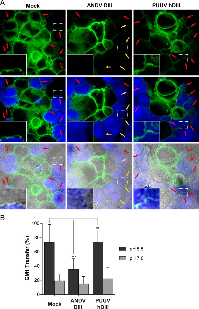
Fig 7. Exogenous DIII and stem fragments block a fusion stage preceding the mixing of outer membrane leaflets.
(A) Fluorescence confocal microscopy of the GM1 transfer from ANDV glycoprotein-expressing 293FT effector cells (GM1+) to CHO-K1 target cells (GM1-), treated at pH 5.5. CHO-K1 cells were stained with CMAC (blue fluorescence) and GM1+ cells were labeled with the cholera toxin β-subunit conjugated to Alexa Fluor 488 (green fluorescence). Red arrows indicate GM1 transfer to CHO-K1 target cells (resulting in GM1+); yellow arrows indicate CHO-K1 target cells negative for GM1 (remaining GM1-). Magnification 60x. (B) Quantification of the GM1 transfer between ANDV glycoprotein-expressing 293FT cells (GM1+, effector cells) to CHO-K1 cells (GM1-, target cells), in the absence (Mock) or presence of recombinant ANDV DIII or PUUV hDIII. CHO-K1 cells were only considered GM1+ when the GM1 signal was detected at the full circumference of the cells. ***, P < 0.00025; **, P < 0.0025; *, P < 0.025; ns, not significant.

