
Figure 1.
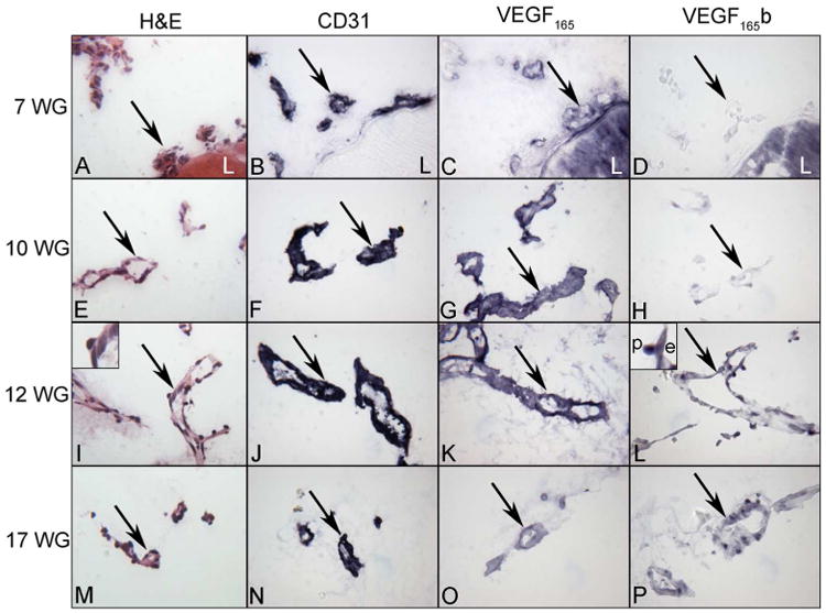
Localization of VEGF165 and VEGF165 b in the human fetal vasculature of vitreous at 7, 10, 12 and 17WG human eyes. Hematoxylin and eosin staining shows the highly cellular and poorly defined lumenal structure of blood vessels at 7 WG which mature into well developed blood vessels by 12 WG. CD31 is expressed until vessels have fully regressed by 21 WG. VEGF165 was present diffusely in blood vessels at 7 WG, increased in staining until 12 WG and decreased by 17 WG. Conversely, VEGF165b was undetectable at 7 WG, increased steadily thereafter, and was associated with nuclei of endothelial cells (e) and pericytes (p) (insets 12 WG). (arrows= blood vessels, L = lens)(APase reaction product)