
Figure 3.
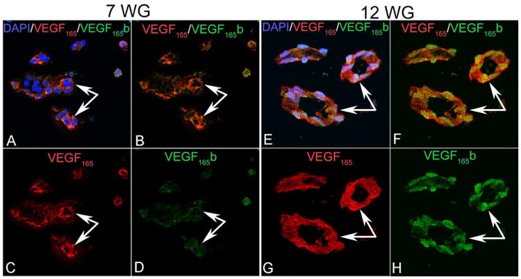
VEGF165 (red) and VEGF165 b (green) in the human fetal vasculature of vitreous at 7 and 12 WG. At 7 WG, moderate VEGF165 immunostaining was present diffusely in blood vessels while very little VEGF165b staining was detected. By 12 WG, VEGF165 immunostaining had increased in endothelial cells and pericytes and VEGF165b labeling was strong and localized to endothelial cell and pericyte nuclei. (arrows indicate blood vessels).