
Figure 6.
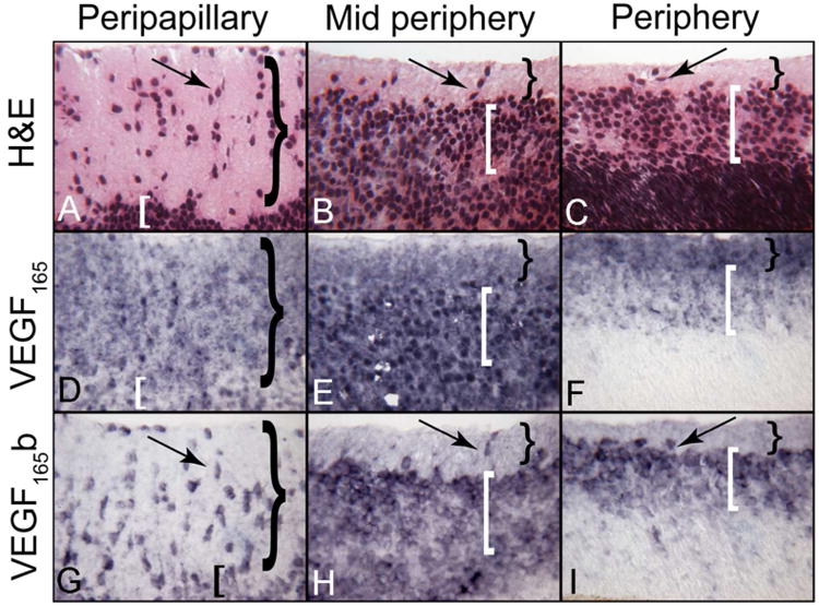
VEGF165 and VEGF165 b localization in different regions of a 12 WG human retina. Structure of developing inner retina at 12 WG showing the nerve fiber layer (NFL= }) and inner neuroblastic layer (INL= [) and cells migrating inwardly (arrows). Both ganglion cells and blood vessels will occupy the inner retina. Regional staining patterns show the diffuse distribution of VEGF165 in both layers while VEGF165b is cell-associated. (APase reaction product)