
FIGURE 1:
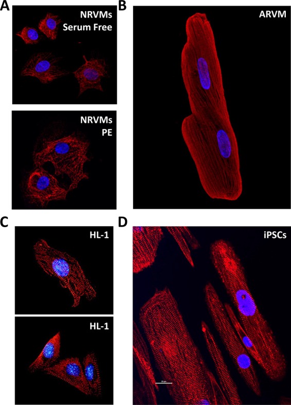
Morphology and sarcomeres in primary cardiac myocytes and the HL-1 cell line. Blue, 4′,6-diamidino-2-phenylindole for nuclei. (A) Mononuclear NRVMs either untreated (top, serum free) or treated with PE (bottom) and stained for myosin heavy chain (red). (B) Binuclear ARVM stained for myosin heavy chain (red). (C) HL-1 cells stained for myosin (top) or titin (bottom). Published with permission from White et al. (2004). (D) iPSC-derived cardiomyocytes cultured on nanopatterned surfaces for 80 to 100 d postdifferentiation induction and stained for α-actinin (image provided by the Michael Regnier laboratory).