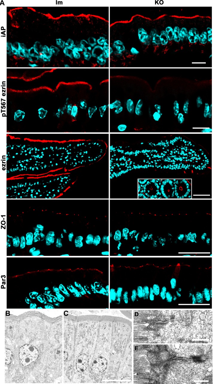
FIGURE 4:
Apical and junction markers and intestinal epithelium ultrastructure in Prkciflox/flox Vil-CRE± (KO) mice. (A) Frozen sections of villus enterocytes from KO and control littermates (lm) were processed with the following antibodies: alkaline phosphatase (iAP), active (pT567) ezrin, ezrin, ZO-1, and human homologue of Par3. Ezrin inset, cross sections of the crypts displaying normal apical ezrin levels. Bars, 10 μm (iAP, pT567ezrin), 45 μm (ezrin), 25 μm (ZO-1, Par3). (B–E) Electron microscopy images of villus enterocytes from control (B, D) or KO (C, E) animals. Bars, 10 μm (B, C), 0.5 μm (D, E).

