FIGURE 3:
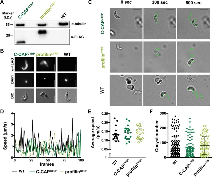
Normal ookinete motility and Anopheles infections in transgenic parasite lines. (A) Western blot analysis of FLAG-tagged proteins in ookinetes. Protein extracts of 100,000 purified ookinetes were separated by SDS–PAGE. Samples were probed against an anti-FLAG antibody and anti-tubulin antibody as control. (B) Immunofluorescence micrographs of ookinetes stained with anti-FLAG antibody and DAPI. Differential interference contrast images are included to display ookinetes (bars, 5 μm). (C) Time-lapse micrographs of representative ookinete at 0, 300, and 600 s. The line reflects ookinete motility in Matrigel (bars, 10 μm). (D) Representative velocities of transgenic and WT ookinetes over a recording period of 500 s. Each frame corresponds to 5 s. (E) Average speed (μm/s) was quantified from time-lapse movies (n = 20 each). The results represent mean values (± SD) of three independent experiments. Differences are nonsignificant (Mann–Whitney and Kruskal–Wallis tests). (F) Oocyst numbers in infected mosquitoes. The results represent mean values (± SD) of five independent natural feeding experiments.

