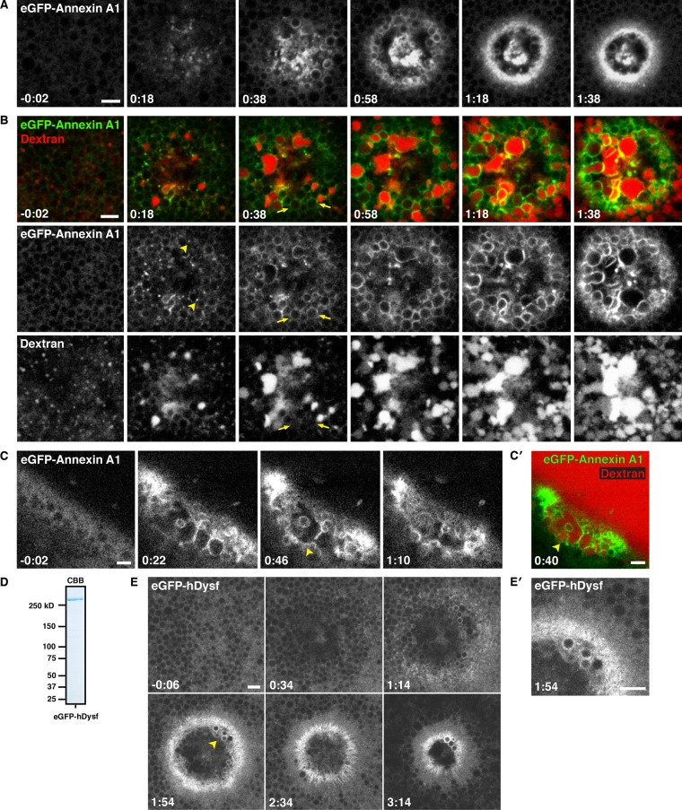
FIGURE 7:
Human annexin A1 and dysferlin are recruited to membranous structures at wounds. (A) Low-magnification, en face view of wounded oocyte expressing eGFP-annexin A1. Annexin is recruited to cortical foci (0:18), vesicles at the wound edge (0:38), and a tight ring around the PM (1:18). See Supplemental Movie S14. (B) En face view of oocyte expressing eGFP-annexin A1 wounded in the presence of fluorescent extracellular dextran (Alexa Fluor 647 dextran). Annexin is recruited to foci (arrowheads) and vesicles before dextran incorporation (arrows). At increasing times postwounding, annexin also labels compartments in the wound interior (1:38). See Supplemental Movie S15. (C) Oblique view of a cell expressing eGFP-annexin A1 wounded in the presence of extracellular fluorescent dextran (not shown), showing eGFP-annexin A1 accumulating at a nascent patch (arrowheads). See Supplemental Movie S16. (C′) Still from C showing that eGFP-annexin A1–labeled compartments (green) fuse to form a barrier that excludes extracellular fluorescent dextran (red; Alexa Fluor 647 dextran) from the cytoplasm. (D) Coomassie-stained SDS–PAGE gel showing recombinant FLAG-eGFP-hDysferlin_isoform 1 purified from Sf9 cells. (E) Oocyte wounded after microinjection with recombinant FLAG-eGFP-hDysferlin. (E′) Enlargement of region indicated by an arrowhead in E, showing recruitment of dysferlin to a tight ring at the PM bordering the wound and to vesicles at wound edge. Time in minutes:seconds, with t = 0:00 corresponding to the moment of wounding. Scale bars, 5 μm.

