. 2016 Jun 28;5:e14692. doi: 10.7554/eLife.14692

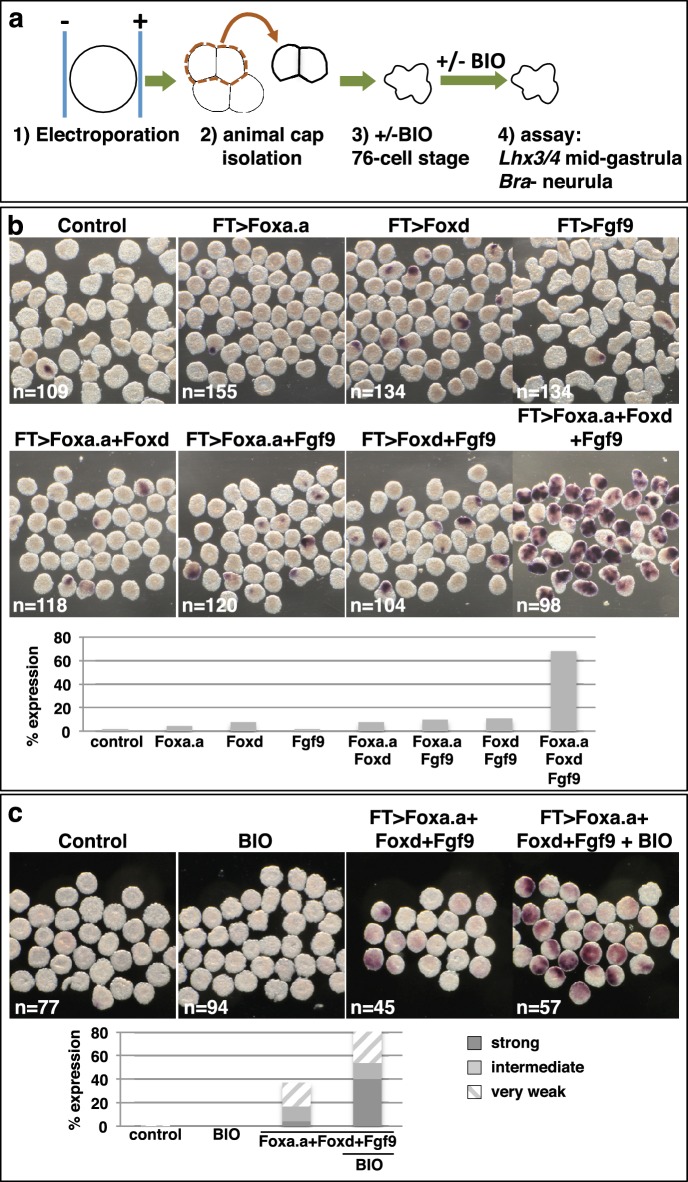
Figure 5. Reprogramming the ectoderm lineage to mesendoderm.

(a) Experimental scheme. Embryos were electroporated and the ectoderm lineage (animal cap) isolated at the eight-cell stage. Ectodermal explants were cultured until the mid-gastrula stage for Lhx3/4 expression or until the neurula stage for Bra expression. Optionally, explants were treated with BIO, when control sibling embryos reached the 76-cell stage, for approximately 1 hr prior to fixation (Lhx3/4 only). (b–c) Expression of Bra (b) and Lhx3/4 (c) in isolated ectodermal explants, following the treatments indicated above the panels. ‘n=’ represents the number of explants analysed. Graphs shows the percentage of explants with any level of Bra expression or level of Lhx3/4 expression indicated by the key, under various conditions (Foxa.a= pFT>Foxa.a; Foxd = pFT>Foxd; Fgf9= pFT>Fgf9/16/20; control = unelectroporated).

DOI: http://dx.doi.org/10.7554/eLife.14692.010

Figure 5.

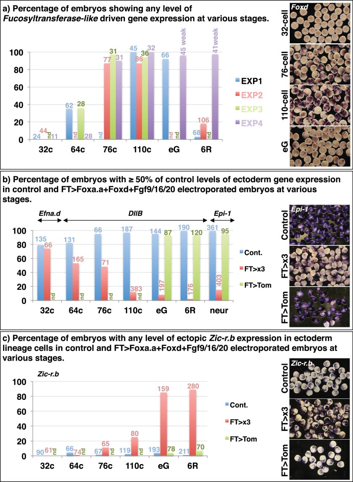
Figure 5—figure supplement 1. Reprogramming of ectoderm cells to mesendoderm fates.

Figure 5—figure supplement 1.

32c=32-cell stage; 64c=64-cell stage; 76c=76-cell stage; 110c=110-cell stage; eG= early gastrula stage (approximately 3-row neural plate stage); 6R=6-row neural plate stage (mid-gastrula); neur = neurula stage. (a) Determining the onset of promoter activity of the Fucosyltransferase-like gene. The pFT>Foxd construct was electroporated and embryos were fixed at different developmental time points and assayed for Foxd expression in ectoderm cells by in situ hybridisation. The graph shows the percentage of embryos showing any level of Foxd expression in ectoderm cells in four independent experiments. The number of embryos counted per bar on the graph is indicated above the bar. nd = not done. On the right are shown examples of Foxd in situ hybridisations on electroporated embryos at the time points indicated. (b—c) FT>x3= pFT>Foxa.a + pFT>Foxd + pFT>Fgf9/16/20; FT>Tom= pFT>tdTomato (as a control electroporation); Cont. = unelectroporated embryos. (b) The ectoderm genetic programme is down-regulated in FT>x3 electroporated embryos. Electroporated embryos were analysed for Efna.d at the 32-cell stage, DllB at the 64-cell to 6-row neural plate stage, and Epi-1 at the neurula stage. The graph shows the percentage of embryos with ectoderm gene expression corresponding to 50% or more of control levels (estimated based on size of expression domain), under the conditions indicated by the key. nd= not done. (c) Zic-r.b is ectopically activated in non-neural ectoderm cells in FT>x3 electroporated embryos. The graph shows the percentage of embryos with any level of ectopic Zic-r.b expression in ectoderm cells, under the conditions indicated by the key. nd = not done.
Figure 5—figure supplement 2. Confirmation that BIO-treatment of ectoderm explants at the 76-cell stage results in nuclear localisation of β-catenin.
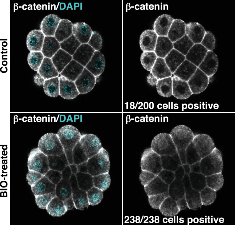
Figure 5—figure supplement 2.

Explants were treated with BIO for 30 minutes and then immunostained with b-catenin antibodies and counterstained with DAPI. Panels show single z-slices of confocal images. Numbers indicate the number of interphase cells with nuclear b-catenin. A total of 11 ectoderm explants were counted for control and 15 for BIO-treated.