
Figure 5. Regulatory features of glycogen synthase.
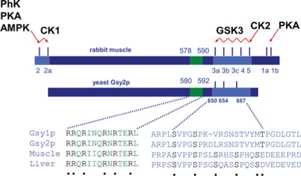
Shown is a comparison of the general architecture of yeast and mammalian glycogen synthases in terms of phosphorylation sites (light blue, not to scale) and the arginine-rich cluster implicated in conferring sensitivity to activation by glucose 6-phosphate (green). The conserved arginine residues and the phosphorylated residues are in black and marked by dots. Some of the protein kinases involved in phosphorylating the mammalian enzyme are linked to sites they modify. See the legend to Table 2.