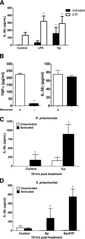
Figure 4. IL-36γ protein detection in conditioned medium of PMs is increased after ATP stimulation or sonication.
(A) WT PMs were treated with LPS (100 ng) or HK Kp (MOI 10:1), with or without the addition of ATP (5 mM), during the last 20 min of incubation. Conditioned medium was collected at 18 h, and IL-36γ protein was assessed by ELISA. Data are means ± sem of 4 wells/group (106 cells/well). *P < 0.05 vs. control. (B) WT PMs were treated with HK Kp (MOI 10:1 for 4 h) and ATP (5 mM for the last 20 min), with or without monensin (25 μM) for the last hour of incubation. Conditioned medium was collected and analyzed for TNFα and IL-36γ by ELISA. Data are means ± sem of 3 samples per group. *P < 0.001 vs. control. (C) WT PMs were treated with or without HK Kp (MOI 10:1). Conditioned medium was collected at 18 h and sonicated (10 s output twice) or not. IL-36γ protein was assessed by ELISA. (D) WT PMs were treated with HK Sp (MOI 10:1), with or without addition of ATP (5 mM) during the last 20 min of incubation. Conditioned medium was collected at 18 h and was sonicated (10s × 2 output) or was not sonicated. IL-36γ protein was assessed by ELISA. (C, D) Data are means ± sem of 3 wells/group (106 cells/well). *P < 0.05 vs. control.

