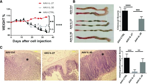
Figure 2. A single dose of AAV-IL-27 but not AAV-IL-30 treatment inhibits the development of autoimmune colitis in mice.
CD45RBhigh T cells were sorted from spleen and lymph node cells from C57BL/6 mice and were injected into Rag1−/− C57BL/6 mice intraperitoneally at a dose of 3 × 10⁵/mouse. At d 12 after T cell transfer, AAV-IL-27, AAV-IL-30, or AAV-ctrl viral vectors were injected into each mouse intramuscularly at a dose of 2 × 10¹¹ DRP/mouse. (A) Mice (n = 4–5/group) were weighted every 3 d and evaluated for the development of wasting disease. (B) Five weeks after T cell transfer, mice were euthanized, and clinical scores of colitis were assigned to each mouse based the macro pathology of the colon, as described in Materials and Methods. (C) Histopathology of each colon tissue samples was analyzed and scored as described in Materials and Methods. Representative micrographs of H&E sections and average scores of each treatment groups are shown. (C, left) *, Areas with severe tissue damage. Data are expressed as means of individual determinations ± se. Statistical analysis was performed using the unpaired Student’s t test. **P < 0.01; ***P < 0.001; ****P < 0.0001.

