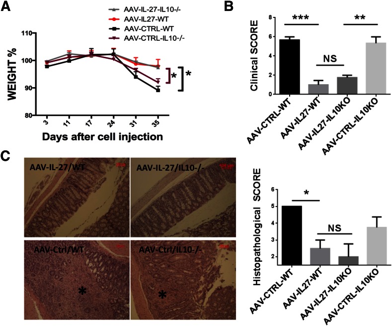
Figure 4. IL-10 is not essential for AAV-IL-27-mediated inhibition of colitis.
(A) Body weight changes following the adoptive transfer of CD4+CD45RBhigh T cells (2.2 × 10⁵/mouse) from WT or IL-10−/− mice. AAV-IL-27 or AAV-ctrl viral vectors were injected intramuscularly into mice (n = 3–6/group), 3 d after T cell transfer. (B) Mice were euthanized, and clinical scores of colitis were assigned to each mouse based on the macro pathology of the colon, as described in Materials and Methods. KO, Knockout. (C) Representative micrographs of H&E sections and average scores of each treatment groups are shown. Longitudinal sections of the entire colon were stained with H&E. Original scale bars, 100 μm. (C, left) *, Area with severe tissue damage. Histopathology scores were calculated as described in Materials and Methods. Data are expressed as means ± se. Statistical analysis was performed using the unpaired Student’s t test. *P < 0.05; **P < 0.01; ***P < 0.001.

