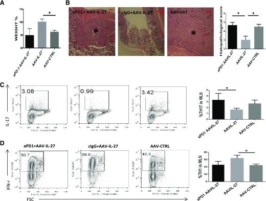
Figure 6. PD-1 blockade breaks T cell tolerance in AAV-IL-27-treated mice.
CD4+CD45RBhigh T cells (5 × 10⁵/mouse) from WT mice were injected into Rag2−/− mice intraperitoneally. Five days after T cell transfer, mice (n = 3–4/group) were treated with AAV-IL-27 or AAV-ctrl viral vectors intramuscularly. On d 34, 37, and 40 after T cell transfer, mice receiving AAV-IL-27 treatment were also treated with 300 μg/mouse of anti-PD-1 (aPD-1) or an isotype-matched ctrl antibody intraperitoneally. Disease development was monitored. (A) Body weight changes at d 42 after T cell transfer are shown. (B) Representative images of histology of colon tissues and average scores from each group of mice are shown. Original scale bars, 100 μm. (B, left) *, Severe tissue damage. cIgG, control IgG. Representative FACS plots of CD4+ T lymphocytes expressing IL-17A (C) and IFN-γ (D) are shown. Three to 4 mice/group were included in this experiment. Statistical analysis was performed using the unpaired Student’s t test. *P < 0.05.

