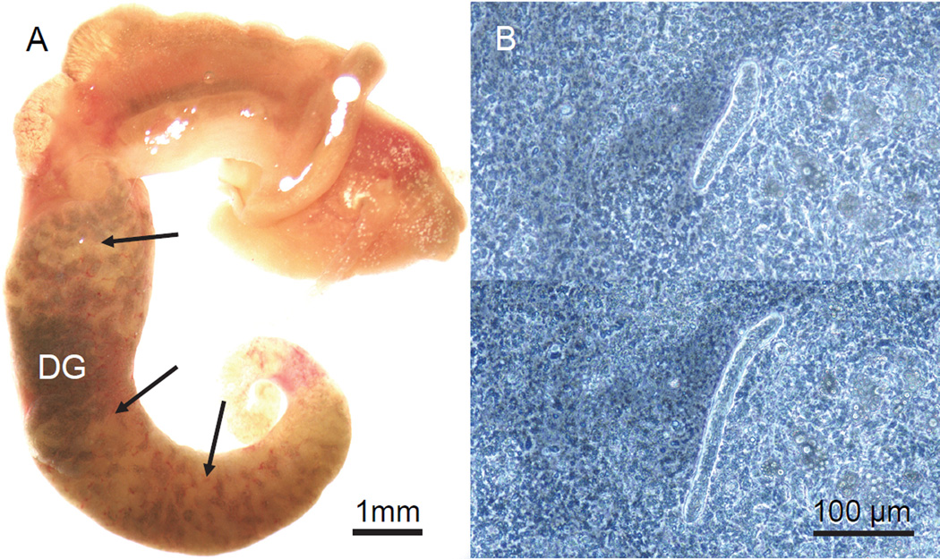
Figure 1.
Sporocysts of Schistosoma mansoni in Biomphalaria glabrata. A. M-line snail dissected from its shell at 21 days post-exposure (DPE) to miracidia. The digestive gland (DG), which occupies most of the posterior half of the snail and is uniformly dark green in uninfected specimens, is heavily infected with cream-colored masses of secondary sporocysts. This snail also was releasing cercariae prior to dissection. B. Phase contrast images of a motile, immature daughter sporocyst in a squash of head foot tissue from a snail at 22 DPE, showing characteristic extension and retraction of the body.

