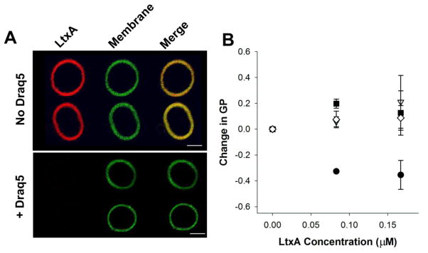
Fig. 3. DRAQ5™-induced changes in LtxA interactions with POPC membranes.
(A) LtxA affinity for POPC membranes. GUVs composed of POPC/Chol, labeled with 1% NBD (green) were pretreated with DRAQ5™ or left untreated. LtxA (red) was then added to the GUVs. LtxA showed a strong affinity for untreated membranes, but binding was abolished in DRAQ5™-pretreated GUVS. Scale bar = 5 μm. (B) LtxA-mediated membrane disruption. The disruption of bilayer packing induced by LtxA was recorded using laurdan fluorescence. POPC liposomes were preincubated with DRAQ5™ at a concentration of 0 μM (black circles), 1 μM (white diamonds), 5 μM (black squares), or 10 μM (white triangles) before addition of LtxA to measure inhibition of LtxA-mediated membrane disruption. A two-way ANOVA indicates that the effect of LtxA depends on the DRAQ5™ concentration (p = 0.005).

