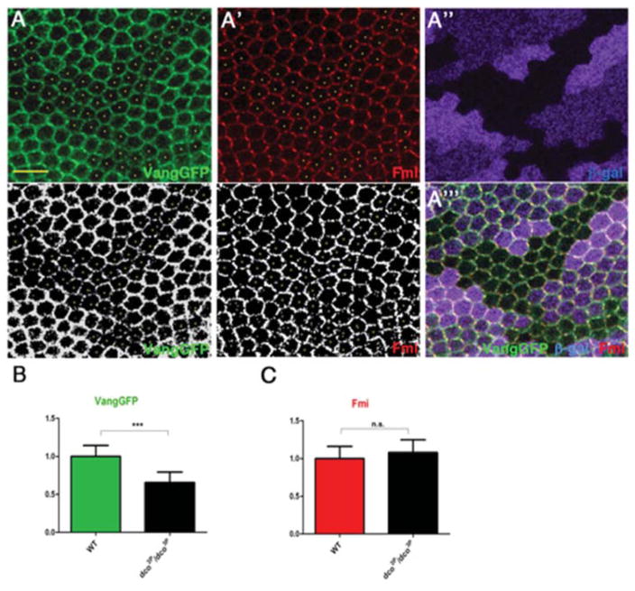
Figure 7. Vang localization is affected in dco mutant clones.
(A–A‴) Confocal images of immunostained pupal wings (at 30hr APF) carrying dco3P clones (marked by absence of β-Gal staining, magenta) and expressing act-VangGFP stained for Vang (green, monochrome in A, bottom panel) and Fmi (red, monochrome in A′, bottom panel). VangGFP levels are reduced in dco3P clones (individual cells marked with yellow dots in A and A′). Scale bar: 10μm.
(B) Mean pixel intensity of cortical VangGFP in dco mutant wing tissue (n=15), normalized to cortical VangGFP mean pixel intensity in wild-type tissue (n=15) (***p<0.001 with student’s t-test, error bars=S.D.).
(C) Mean pixel intensity of cortical Fmi in dco mutant wing tissue (n=12 cells), normalized to cortical Fmi mean pixel intensity in wild-type tissue (n=12 cells) (n=5); n.s. p>0.05 with student’s t-test (error bars=S.D.).

