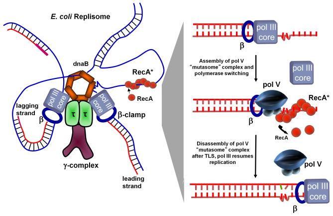
Fig. 4.
TLS cis-activation model. A model of an E. coli replisome stalled at a lesion site is depicted in the left panel and individual TLS steps are shown in the right panel. DNA pol III stalls at the lesion site, DnaB helicase unwinds dsDNA ahead of the blocked fork resulting in regions of ssDNA where RecA* assembles in cis. The cis-activation model requires polymerase switching. DNA pol V replaces pol III core on the β clamp and bypasses lesions with concurrent 3′ → 5′ displacement of RecA* formed ahead of the lesion by pol V. Following TLS, pol III core replaces pol V to continue rapid replication of undamaged DNA downstream of the lesion.

