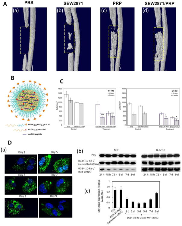
Figure 3. Examples of controlled release strategies used to modulate the M1-M2 balance in tissue engineering applications.

A) Use of a hydrogel scaffold loaded with immunomodulators. Three-dimensional microCT images of bone regeneration in a rat defect model six weeks after implantation of hydrogels loaded with (a) PBS, (b) SEW281, (c) PRP, and (d) SEW287 and PRP are shown. Reprinted with permission from Kim et al., 2014 [66]. (B) Encapsulation of N-terminal peptide consisting of amino acids 2-26 of Annexin A1 (Ac2-26) in a PLGA-PEG nanoparticle conjugated to a peptide targeting collagen IV. Reprinted with permission from Kamaly et al. (2013) [75]. (C) Decellularized small intestinal submucosa (SIS) was coated with anti-inflammatory peptide amphiphiles (AIF-PAs) derived from uteroglobin protein sequences. Five weeks after implantation in a rat bladder augmentation model, SIS coated with peptides 1 or 2 (SIS/AIF-PA1/2) showed decreased numbers of M1 macrophages (labeled as M1+M2- where CD86+ cells were labeled as M1+) compared to uncoated (SIS) scaffolds or scaffolds coated with a control peptide amphiphile (SIS/AIFC-PA6). After five weeks, SIS/AIF-PA1/2 scaffolds had lower total numbers of M1 and M2 cells (M1+M2+ where CD206+ were labeled as M2+) compared to control scaffolds. Top and bottom row of asterisks represent comparisons to SIS and SIS/AIFC-PA6, respectively, and * represents p < 0.05, ** represents p < 0.01, *** represents p < 0.001, and **** represents p < 0.0001. Reprinted from Bury et al. (2014), with permission from Elsevier [81]. (D) Delivery of AF488-siRNA and MIF expression within primary macrophages. (a) Confocal microscopy images of macrophage cultures. Nuclei were stained with DAPI (blue), and nanoparticles with siRNA were dyed green. (b) Western blots showing MIF and β-actin (used for normalization) protein expression in macrophages. (c) qRT-PCR analysis of MIF gene expression in macrophages. PBS and scrambled siRNA served as negative controls. Reprinted with permission from Zhang et al., 2015 [89].
