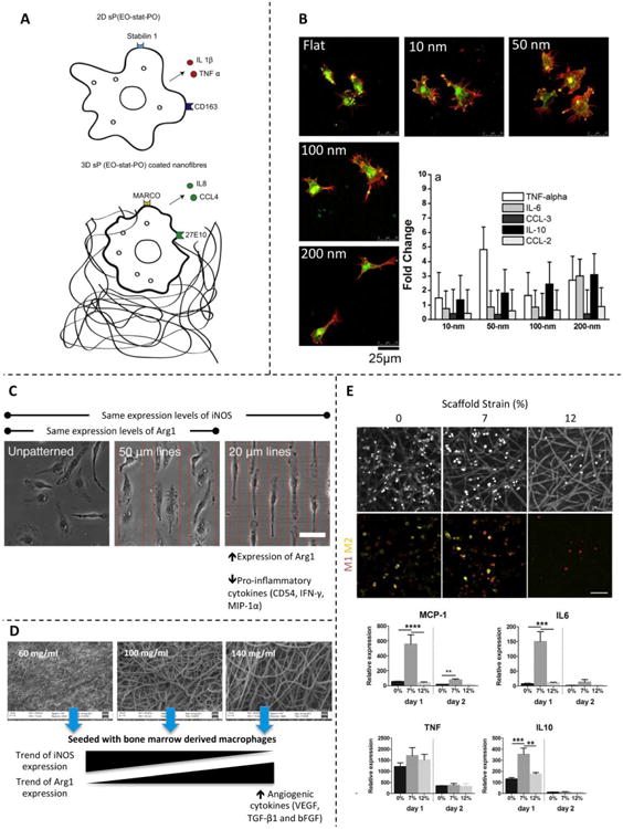
Figure 4. Effect of physical cues found in different scaffolds.

(A) Cartoon showing the effect of culturing macrophages in 2D vs. 3D substrates of the same chemical composition. Reprinted with permission from Bartneck et al., 2012 [101]. (B) Macrophages seeded in scaffolds with nanotopographies composed of nanodots on the order of 10–200 nm. Micrographs show the effect on cell shape, and histograms show the effect on the cytokine and chemokine gene expression profile. Reprinted with permission from Mohiuddin et al., 2012 [98]. (C) Micropatterned scaffolds promote cell elongation. The induced cell shape derives from expression of differential markers and secretion of several cytokines. Reprinted with permission from McWhorter et al., 2013 [32]. (D) Electrospun sheets containing various polymer concentrations form scaffolds with different porosities and surface area. Macrophages respond differently to each scaffold architecture and show M2-like behavior when seeded on scaffolds produced with high concentrations of polymer. Reprinted with permission from Garg et al., 2013 [102]. (E) Macrophages adhering to electrospun scaffolds and subjected to different strain levels. The applied deformation affected the macrophage phenotype and gene expression profile. Reprinted with permission from Ballota et al., 2014 [109].
