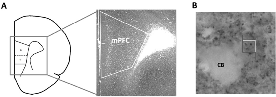
Fig. 1.
Coronal section of the mPFC that illustrates the immuno-labeled tissue used for counting synaptophysin across all cellular layers. PL = prelimbic, IL = infralimbic (A). Photograph within the mPFC showing immunoreactive synaptophysin boutons. The stereological counting frame (4µm × 4µm) is shown. CB = cell body (B).

