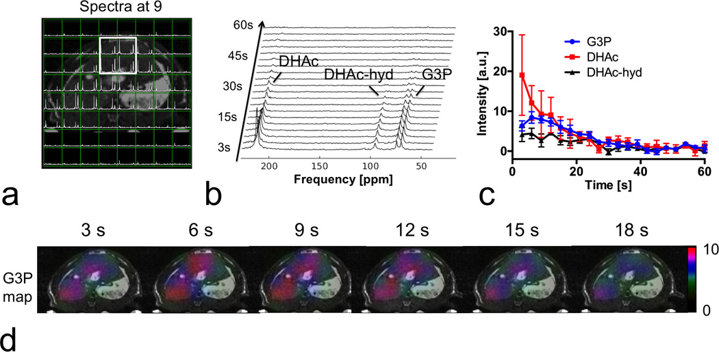
Figure 6.

Prospectively 3.8-fold accelerated CS MRSI on rat liver, following intravenous administration of hyperpolarized [2-13C] dihydroxyacetone (DHAc), which results in a 140-ppm (4.5 kHz at 3T) range of metabolite chemical shifts. (a) A mosaic view of spectra and abdominal T2-weighted MRI. Spectra were spatially localized primarily within the liver as expected. (b) Dynamic spectra from the marked area in (a). The three peaks in the dynamic spectra are DHAc (≤0.3° flip, 213 ppm), DHAc hydrate (DHAc-hyd, 2.3° flip, 96 ppm) and glycerol 3-phosphate (G3P, 20° flip, 73 ppm), all of which were recovered by the proposed method. (c) Time courses of DHAc, DHA-hyd and G3P from the marked liver area. (d) The 13C metabolic map showed that G3P generated from DHAc was primarily distributed within the liver, indicating accurate reconstruction by our method.
