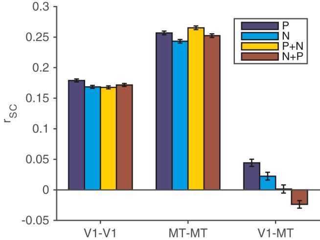
Figure 2.

Spike count correlations between pairs of units in different, but not the same, cortical areas depend on the visual stimulus. We compared rSC between units with similar direction tuning in four stimulus conditions: a single stimulus moving in the preferred direction (dark blue bars), a single stimulus moving in the null or antipreferred direction (light blue bars), a preferred stimulus in the V1 unit's receptive field combined with a null stimulus outside the V1 unit's receptive field (but still inside the MT neuron's receptive field; yellow bars), and a null stimulus in the V1 unit's receptive field combined with a preferred stimulus outside the V1 unit's receptive field (red bars). Spike count correlations did not substantially depend on the visual stimulus for pairs of V1 units (left bars) or pairs of MT units (middle bars), but they did depend on the stimulus for pairs of V1 and MT units (right bars; t tests on rate-matched distributions, p < 0.05; for detailed statistics, see Data analysis). Error bars represent SEM.
