
Figure 4. Influence of rapidly activating potassium current (IKr) on repolarization in the human failing heart: action potential duration reduction (ΔAPD) values with IKr blockade .
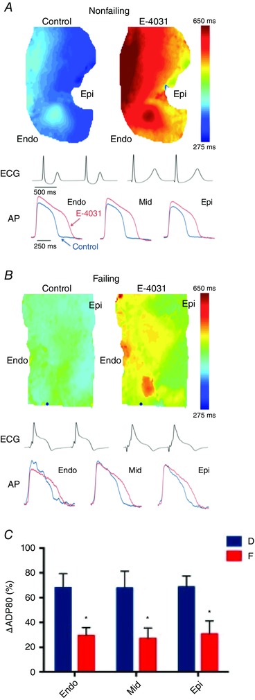
A and B, representative action potential duration (APD) maps, optical recording traces, and ECGs for donor (non‐failing, A) and failing (B) hearts. C, ΔAPDs, expressed as a 1% control for 1000 ms pacing cycle lengths (PCLs). Data are expressed as means ± SEM. Adapted from Holzem et al. (2015).