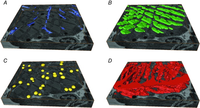
Figure 6. Ultrastructure nano‐imaging of a human cardiomyocyte .

The sectioned volume is 14.7 μm × 14.8 μm × 2.2 μm and the cardiomyocyte was sectioned in 10 nm slices. A, transverse tubules (blue). B, A‐band (green). C, lipid droplets (yellow). D, mitochondria (red). Adapted from Sulkin et al. (2014).
