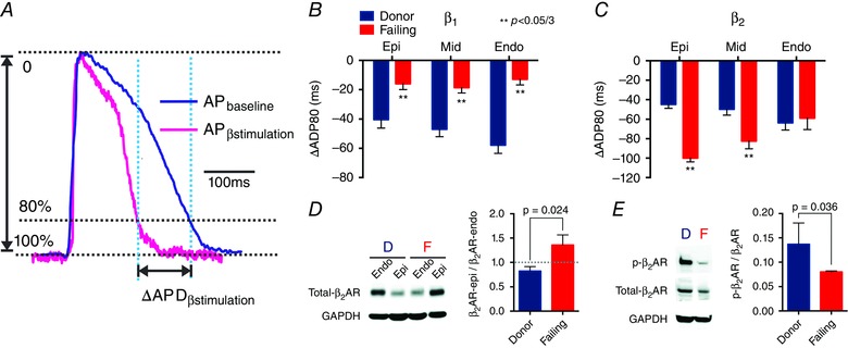
Figure 8. Effect of β‐adrenergic receptor (β‐AR) stimulation on repolarization, protein expression, and protein phosphorylation .

A, AP signals illustrate that the difference between APDbaseline − APDβ‐stimulation is ΔAPD. B, statistical analysis of ΔAPD shows desensitization of β1‐AR at the sub‐EPI, MID, and sub‐ENDO. C, statistical analysis of ΔAPD shows sensitization of β2‐AR at the sub‐EPI and MID but not at the sub‐ENDO. B and C, negative values indicate a reduction in APD. D, Western blot reveals a reversal of the sub‐EPI to sub‐ENDO gradient of β2‐AR protein expression (β2‐AR‐epi/β2‐AR‐endo) in HF. E, phosphorylation of β2‐AR is reduced in HF. D, donor hearts; F, failing hearts. Adapted from Lang et al. (2015).
