Figure 1.
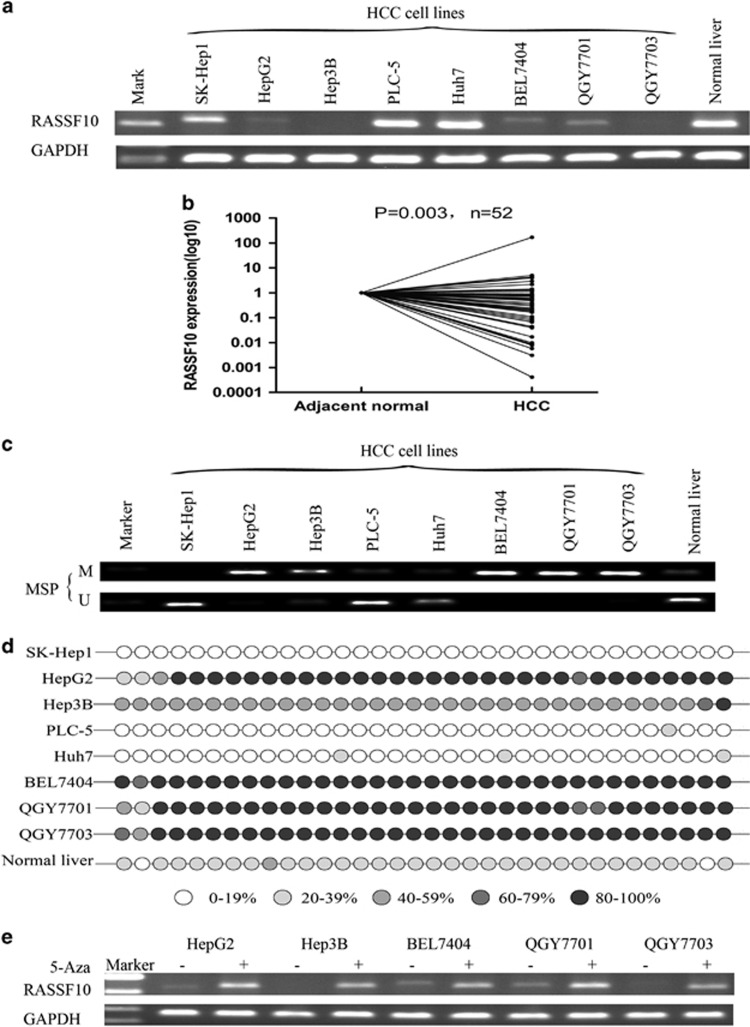
The expression of RASSF10 and its promoter methylation status. (a) The expression profile of RASSF10 mRNA in HCC cell lines and normal liver tissue by RT-PCR. (b)The mRNA expression level of RASSF10 was significantly down-regulated in primary HCCs as compared with their adjacent normal tissues by quantitative real-time PCR (P=0.003, n=52). RASSF10 expression level was normalized with the GAPDH mRNA level. (c) Methylation-specific polymerase chain reaction showed methylation of RASSF10 in HCC cell lines. M, methylated DNA; U, unmethylated DNA. (d) BGS confirmed the methylation status of RASSF10 in HCC cell lines and normal control. (e) RASSF10 mRNA expression was restored following 5-aza-DC treatment. GAPDH was used as a control for equal loading.

