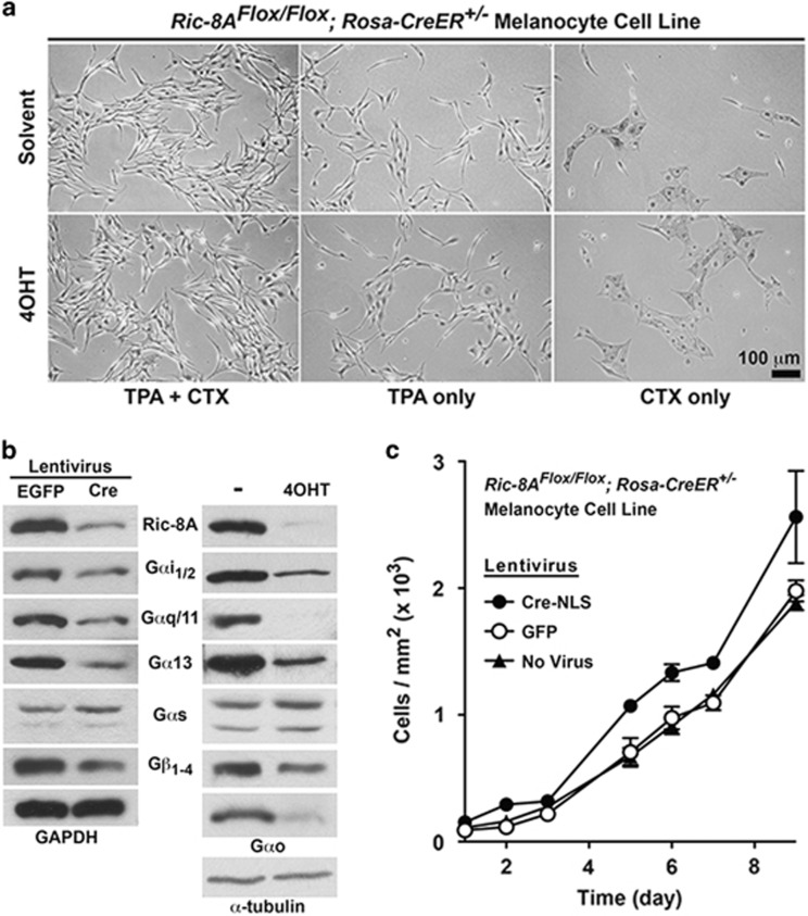
Figure 1.
Deletion of Ric-8A in murine melanocytes confers a modest cell proliferation advantage, but does not confer TPA- and/or CTX-independent growth. (a) Bright-field images of untreated or 4OHT-treated immortalized Ric-8AFlox/Flox; Rosa-CreER+/− melanocytes grown in the presence of CTX, or TPA, or both for 4 days. (b) Quantitative western blot analyses of Ric-8A, Gαi1/2, Gαq/11, Gα13, Gαs, Gαo and Gβ1−4 levels in Cre or GFP (control) lentivirus, or 4OHT-treated and untreated cultured melanocyte cell lines. Relative GAPDH or α-tubulin levels are shown as normalization controls. (c) Cell proliferation analyses of untransduced, and Cre or GFP (control) lentivirus-infected Ric-8AFlox/Flox; Rosa-CreER+/− melanocytes. Error bars are the mean±s.e.m. of experiments performed in triplicate.

