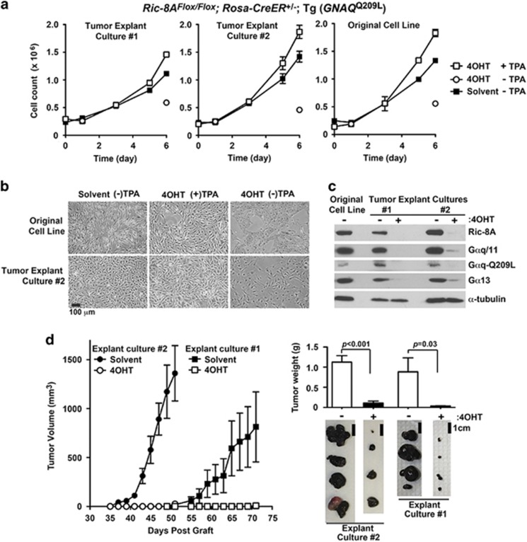
Figure 4.
GNAQQ209L-driven secondary melanoma tumor progression is attenuated by induced Ric-8A deletion. (a) Two explanted GNAQQ209L tumors from Figure 3a were cultured ex vivo in standard melanocyte culture medium lacking TPA to establish Ric-8AFlox/Flox; Rosa-CreER+/−; (Tg) GNAQQ209L melanoma cell lines. The growth kinetics of the melanoma cell lines±TPA supplementation and ±4OHT to induce ex vivo Ric-8A deletion as indicated were compared with the growth progression of the original Ric-8AFlox/Flox; Rosa-CreER+/−; (Tg) GNAQQ209L melanocyte cell line. (b) Bright-field images of the parental Ric-8AFlox/Flox; Rosa-CreER+/−; (Tg) GNAQQ209L melanocyte cell line and melanoma explant culture #2 at the conclusion of the 6-day growth study. Error bars are the mean±s.e.m. of experiments performed in triplicate. (c) Quantitative western blot analyses of Ric-8A, Gαq/11, Gαq-Q209L and Gα13 levels in lysates prepared from the parental Ric-8AFlox/Flox; Rosa-CreER+/−; (Tg) GNAQQ209L melanocyte cell line and melanoma cell cultures derived from the two independent explanted GNAQQ209L-melanoma tumors. Tumor cell cultures were treated with 4OHT to induce Ric-8A knockout ex vivo. α-Tubulin levels were measured as a normalization control. (d) Secondary tumor progression from grafted Ric-8AFlox/Flox; Rosa-CreER+/−; (Tg) GNAQQ209L explant cultures #1 and #2 after in vitro treatment with solvent or 4OHT to induce Ric-8A deletion. Shown alongside are images and average weights of harvested secondary tumors at day 56 (culture #2) and day 71 (culture #1). Error bars are the mean±s.e.m. (n=4 tumors).

