Figure 2.
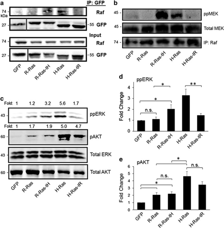
Ras targeting domains dictate access to Raf and MAPK signaling. (a) NIH3T3 murine fibroblasts were stably transfected with GFP-tagged Ras variants as indicated, and GFP fusion proteins were immunoprecipitated (IP) from cell lysates (Input) with α-GFP antibodies, followed by immunoblotting with α-Raf or α-GFP antibodies. (b) Raf kinase assay. Cells were serum-starved, and Raf activity was assessed as described in Materials and methods. Immunoblotting of the IP fraction with α-Raf antibodies is shown in the lower panel. (c) ERK and AKT activation in Ras-expressing cells after 72 h serum starvation, as assessed by immunoblotting of cell lysates with the indicated antibodies. Phospho:total ratios are shown above the respective blots as a ratio to GFP. (d, e) Fold change in phospho-ERK:total ERK or phospho-AKT:total AKT ratios compared with GFP control+s.e.m. *P<0.05; **P<0.003, n.s., not significant. All blots representative of five independent experiments.

