
Fig. 2.
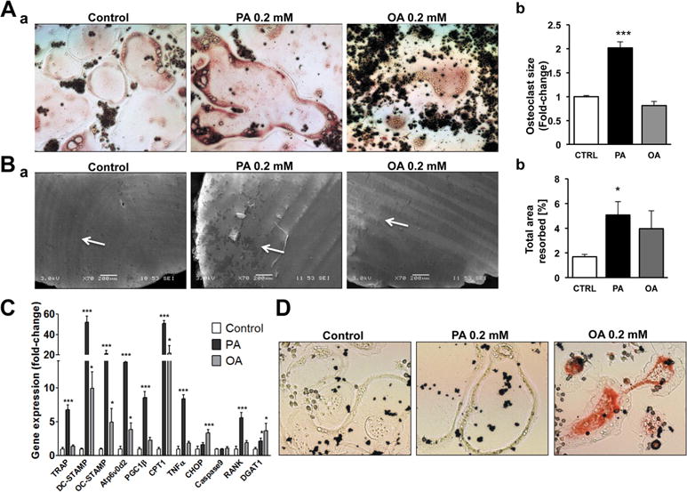
PA enhances osteoclastogenesis. (A) a) TRAP staining in osteoclasts (5 days post- stimulation with RANKL) treated with PA or OA. b) Average size of osteoclasts treated with PA or OA. Average size was calculated from measurement of 233 osteoclasts in control treatment, 202 osteoclasts in PA-treated cultures, and 160 osteoclasts in OA-treated cultures; ***p < 0.001. (B) a) Analysis of osteoclastic resorption using scanning electron microscopy. Cells were cultured in the presence of PA or OA on dentin discs. b) Resorbed area in cultures treated with PA or OA versus CTRL; PA versus CTRL; *p < 0.05, n = 7 to 10. (C) RT-PCR for TRAP, DC-STAMP, OC-STAMP, Atp6v0d2, PGC1β, CPT1, TNFα, CHOP, Caspase9, RANK, and DGAT1 mRNAs; *p < 0.05, ***p < 0.001, n = 4 to 6. D. Neutral lipid staining with Oil-Red-O in osteoclasts treated with PA or OA.