Fig. 6.

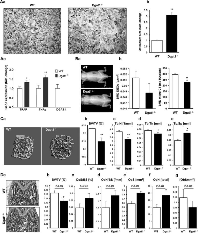
Osteoclastogenesis is increased and femoral bone volume is decreased in Dgat1−/− mice. (A) a) TRAP staining; b) average size of osteoclasts (*p < 0.05; average osteoclast size was calculated from measurement of 311 WT osteoclasts and 267 Dgat1−/− osteoclasts); and c) RT-PCR analysis for TRAP, TNFα, and DGAT1 mRNAs derived from bone marrow cells from 20-week-old Dgat1−/− mice and WT mice; *p < 0.05, **p < 0.01, n=6 to 9. (B) a) DXA scans of 12-week-old Dgat1−/− and WT mice. b, c) BMD of Dgat1−/− and WT mice measured from DXA scan (b, n = 7 to 8) and micro-CT (c, femur trabecular bone, n = 6 to 7, *p < 0.01). (C) a) 3D reconstruction of femoral trabecular bone in 12-week-old Dgat1−/− and WT mice. b–e) Morphometric indices calculated from micro-CT analysis of femurs in 12-week-old Dgat1−/− and WT mice, BV/TV (b), Tb.N (c), Tb.Th (d), and Tb.Sp (e). *p < 0.05, **p < 0.01, ***p < 0.001, n = 6 to 8. (D) Histomorphometric analysis. a) Longitudinal histological sections of femurs of Dgat1−/− and WT mice. b–g) Morphometric indices calculated from histomorphometric analysis of femurs derived from 12-week-old Dgat1−/− and wild-type mice. BV/TV (b), OcS/BS (c), OcN/BS (d), OCS (e), OCN (f), ObS (g). *p < 0.05, n = 5.
