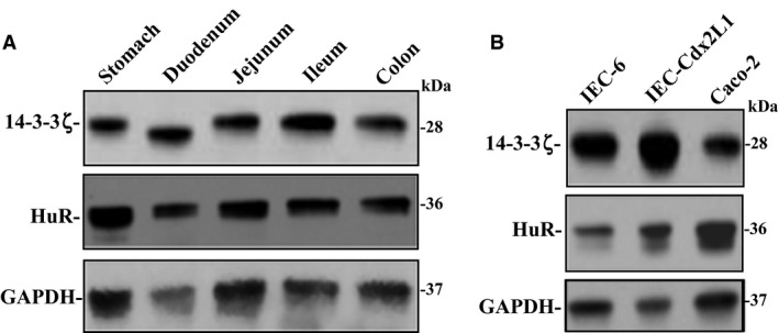
Figure 1.

Levels of 14‐3‐3ζ and HuR in gut mucosa in mice and different lines of cultured IECs. (A) Representative immunoblots of 14‐3‐3ζ and HuR in the mucosa isolated from stomach, duodenum, jejunum, ileum, and colon as examined by western blot analysis. GAPDH immunoblotting was performed as an internal control for equal loading. (B) Representative immunoblots of 14‐3‐3ζ and HuR in IEC‐6, differentiated IEC‐Cdx2L1, and Caco‐2 cells. Three separate experiments were performed that showed similar results. IECs, intestinal epithelial cells.
