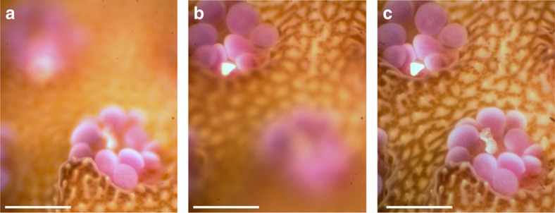
Figure 2. Focal scan using ETL and composite image formation.
Images of a live coral acquired in situ with the BUM using an ETL focal scan. Images collected using the × 5 objective and wide spectrum white LED illumination. (a) Image of a single focal plane showing only the front coral polyp in focus. (b) Image of a single focal plane showing only the back coral polyp in focus. (c) A composite focus stacked image formed using the in-focus portions of 20 images collected with the ETL focal scan. Within a single frame (a,b), the microscope objective yields a shallow DOF. However, the composite focus stacked image, as shown in c, combines frames to provide an enhanced DOF such that both polyps and surrounding area are all in focus. Scale bars, 500 μm.

