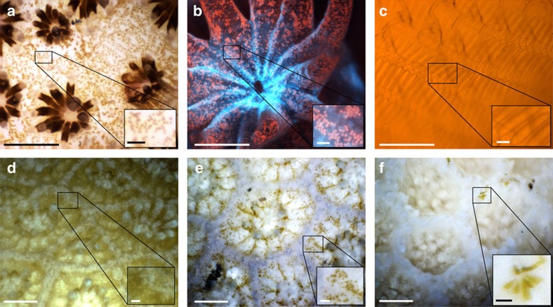
Figure 3. Images captured by the BUM.
(a) In situ image of the coral Stylophora taken using the × 5 objective lens and white illumination, in Eilat, Israel. The image is an enhanced DOF composite formed from a focus stack. Individual zooxanthellae (∼6–13 μm in size) are visible in the inset. (b) Fluorescent image of the coral Pocillopora taken in a lab tank using the × 5 objective. Inset shows individual zooxanthellae emitting red fluorescence from their chlorophyll. Image is a composite focus stack. (c) In situ image of the pharyngeal basket of the semi-transparent ascidian Rhopalaea idoneta, taken using the × 5 objective and white illumination, in Eilat, Israel. R. idoneta is a filter feeder that uses the mesh of the pharyngeal basket to capture plankton. (The orange colour here is likely due to a subject behind the ascidian). (d,e) In situ images of two different locations on a bleaching colony of Porites compressa, taken in Maui, Hawaii. Images show partial bleaching in d and nearly complete bleaching in e. Images are composite focus stacks collected using the × 3 objective lens. (f) In situ image of a fully bleached colony of Porites lobata. Taken in Maui, Hawaii with the × 3 lens. No visible zooxanthellae can be seen in the coral; as a result the polyps have a translucent appearance. While translucent, the polyp structure and tentacles remain intact and visible, indicating that the polyp is still alive. Coenosarc tissue normally connecting polyps is either very thin, or may have fully retracted towards the polyps' centres exposing the coral skeleton. Inset shows colonization of the area between two live polyps by benthic diatoms. Main figure scale bars, 500 μm. Inset scale bars, 50 μm.

