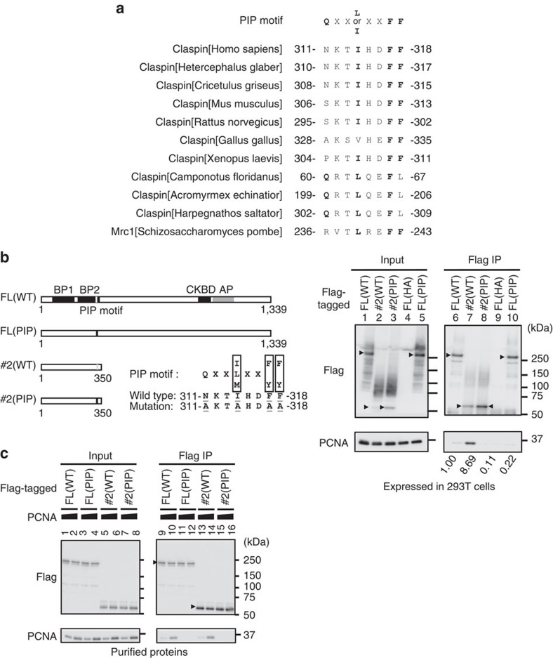
Figure 5. PIP-dependent binding of PCNA to Claspin.
(a) Comparison of the putative PIP motif sequences of Claspin homologues from various species. Conserved amino acids are in bold. (b) Left, a schematic diagram of the full-length and #2 polypeptide of Claspin. Grey and black vertical bars indicate the wild-type and mutant PIP, respectively. Right panels, the wild-type (WT) and PIP mutant (PIP) proteins expressed in 293T cells were pulled down with M2 Flag beads, and analysed by western blotting using anti-Flag or anti-PCNA antibody. FL(HA), HA-tagged wild-type Claspin as a negative control. Arrowheads indicate the full-length and #2 Claspin polypeptides. Values under lanes 6, 7, 8 and 10 represent relative intensities of the coimmunoprecipitated PCNA bands. (c) Interaction between purified Claspin polypeptides and PCNA. PCNA, 1.2 or 3.0 pmol; full-length or #2 Claspin polypeptide, 1.2 pmol each. Pulled down materials by Dynabeads-conjugated anti-Flag antibody were analysed by western blotting using anti-Flag or anti-PCNA antibody.

