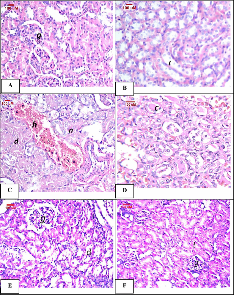
Figure 4.
Representative photomicrographs of kidney sections stained by H&E: (A,B) Control group showing normal glomeruli (g) and normal histological structure of the tubules (t). (C,D) Cisplatin-injected group showing necrosis (n), degeneration (d) and cystic dilatation of the tubules with focal hemorrhages (h) in between. Besides, homogenous eosinophilic casts in lumen of medullary tubules (c) were shown. (E) Indole-3-carbinol pre-treated rats showing degeneration (d) in the lining epithelium of the tubules. (F) Indole-3-carbinol only-treated rats showing normal histological structures.

