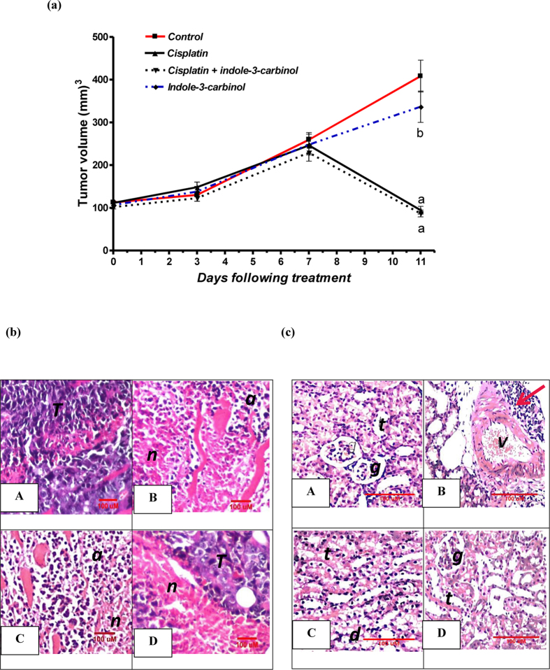
Figure 6.
(A) Effects of cisplatin and/or indole-3-carbinol treatment on tumor growth in Ehrlich ascites carcinoma solid tumor model in mice. a or b: Statistically significant when compared to the control or the cisplatin group, respectively, P < 0.05 using ANOVA followed by Tukey-Kramer as post-hoc test. (B) Representative photomicrographs of Ehrlich ascites carcinoma solid tumor sections taken from the different treatment group and stained by H&E. T: intact structure of tumor cells. n: necrosis of tumor cells. a: apoptosis of tumor cells. (C) Representative photomicrographs of H&E-stained kidney sections taken from the different groups in Ehrlich ascites carcinoma solid tumor model in mice. A: Untreated mouse bearing Ehrlich ascites carcinoma solid tumor showing normal glomeruli (g) and tubules (t) at the renal cortex. B: Ehrlich ascites carcinoma solid tumor-bearing mouse injected with cisplatin (5 mg/kg) showing degeneration together with focal aggregation of lymphoid cells (red arrow) surrounding the congested blood vessel (v). (C) Ehrlich ascites carcinoma solid tumor-bearing mouse treated with cisplatin and indole-3-carbinol showing degeneration (d) in the epithelial lining of the tubules (t). (D) Ehrlich ascites carcinoma solid tumor-bearing mouse treated with indole-3-carbinol alone showing normal glomeruli (g) and tubules (t).

