
Figure 1.
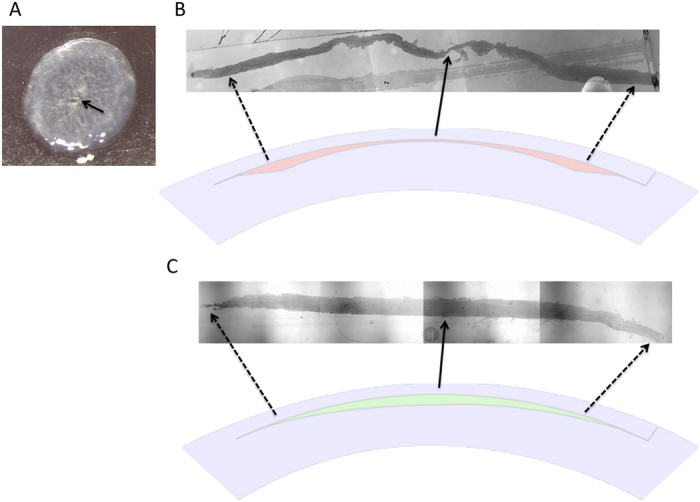
Picture of a button-hole (arrow) in the extracted hyperopic lenticule (A). Semi-thin tissue sections with 1% Fuchsin red staining showing the shape of an extracted +4.00 D hyperopic lenticule (B) and an extracted −4.00 D myopic lenticule (C) for comparison. The extracted hyperopic lenticule was thinnest at the central part and gradually became thicker from the center to periphery (B). Original magnification: 50x. The illustrations show the corneal cross section in the hyperopic-SMILE and myopic-SMILE procedures.