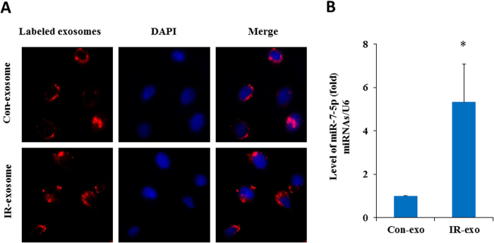
Figure 4. Exogenous exosomes uptake and detection of miR-7-5p in the recipient cells.
Panel A: BEP2D cells were irradiated with or without 2 Gy of 60Co γ-rays, 4 hr later, the exosomes were collected from the medium of cells cultures. The exosomes were labeled with CM-Dil fluorescent dye, and then added to the culture of non-irradiated BEP2D cells. The exosomes uptake was observed by the fluorescent staining. Nuclei were stained blue with DAPI, while exosomes were stained red. Panel B: RT-qPCR estimated the level of miR-7-5p in the non-irradiated cells upon uptake of exogenous exosomes from irradiated cells (IR-exo) or non-irradiated cells (Con-exo). *p < 0.01 as compared with the cells treated with the control exosomes (con-exo) from non-irradiated cells.

