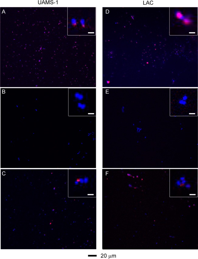
Figure 7.

Two-photon fluorescence images of S. aureus cells treated at different conditions: (A) UAMS-1 exposed to AuNC@PDA–aSpa; (B) UAMS-1 exposed to AuNC@PDA; (C) UAMS-1 spa mutant exposed to AuNC@PDA–aSpa; (D) LAC exposed to AuNC@PDA–aSpa; (E) LAC treated with AuNC@PDA; (F) LAC spa mutant exposed to AuNC@PDA–aSpa. Cells were stained with DAPI colored in blue. Luminescence of AuNCs was colored in red.
