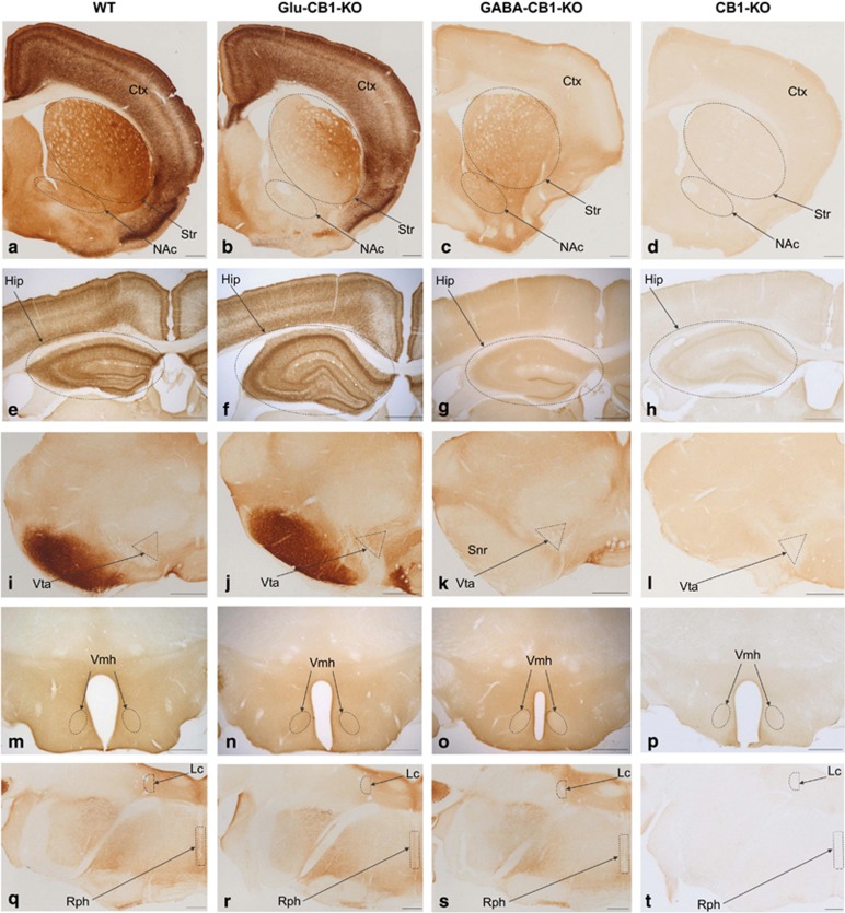
Figure 1.
CB1 protein brain expression in WT, Glu-CB1-KO, GABA-CB1-KO and CB1-KO mice. Cortex (in a–d), striatum (striped oval in a–d), nucleus accumbens (striped oval in a–d), hippocampus (striped oval in e–h), ventral tegmental area (striped triangle in i–l), substantia nigra pars reticulata (in i-l), ventromedial nucleus of the hypothalamus (striped oval in m–p), raphe nuclei (striped rectangle in q–t) and locus coeruleus (striped semi-circle in q–t). Only unspecific diaminobenzidine background is detected in CB1-KO tissue. Relative to WT mice, the Glu-CB1-KO showed a mild decrease in CB1 immunoreactivity while the GABA-CB1-KO showed a more pronounced decrease. CB1 labeling in dorsomedial and ventral striatum is reduced in Glu-CB1-KO (b), whereas substantia nigra pars reticulata lacks CB1 staining in GABA-CB1-KO (k). Note the typical strong CB1 pattern in the inner 1/3 of the dentate molecular layer of GABA-CB1-KO (g). Ctx, Cortex; Hip, Hippocampus; Lc, locus coeruleus; NAc, nucleus accumbens; Rph, raphe nuclei; Snr, substantia nigra pars reticulata; Str, Striatum; Vmh, ventromedial nucleus of hypothalamus; Vta, ventral tegmental area. Scale bars: 500 μm.

