
Figure 3. The recruitment of ribosomes to highly expressed genes does not contribute to domain boundary formation.
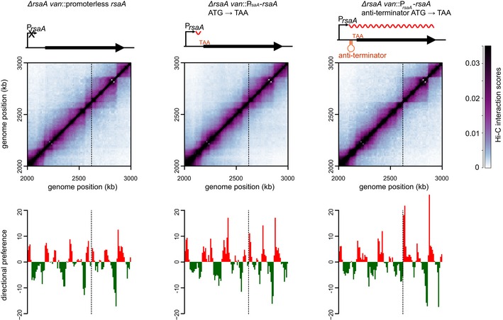
Normalized Hi‐C contact maps for ΔrsaA van::promoterless rsaA, ΔrsaA van::PrsaA‐rsaA (ATG→TAA), and ΔrsaA van::PrsaA‐rsaA (anti‐termination + ATG→TAA) cells. Only the region of the genome containing the van locus (dashed line) is shown. Above each Hi‐C contact map is a schematic of the corresponding rsaA construct. The transcription anti‐terminator is shown as a red hairpin, the start codon mutated to TAA is in red font, and the transcript produced is shown as a wavy red line. Below each map is the corresponding directional preference plot.