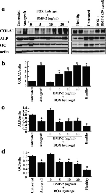
Fig. 10.

Expression of bone regeneration markers in femoral defects after treatment. a After 12-week treatments, the indicated samples were examined for the expression of COL1A1, ALP and OC by Western blot analysis. After normalization with the β-actin content, the relative expression levels of COL1A1, ALP and OC were shown in (b, c and d). Healthy: healthy femora, *P < 0.05 compared to the untreated group
