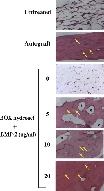
Fig. 9.

Histological analysis (H&E stain, 200x) of bone regeneration in femoral defect rabbits after treatment. The tissue sections from the mid-defect femur regions of rabbits were prepared and stained with hematoxylin and eosin. Homogenous red staining: bone collagen; arrows: osteocyte; asterisks: femoral bone cavities
