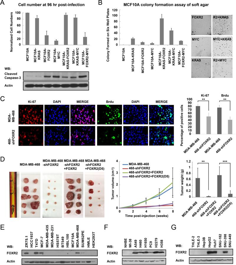
Figure 4. FOXR2 facilitates MYC's activities and promotes tumor proliferation both in vitro and in vivo.
(A) MCF10A cells were infected in six-well plates with retroviruses encoding RAS, MYC, FOXR2, or any two together. Cells were counted after 96 hours of infection. Normalized cell numbers are presented. Data are averages (± SD) of three independent experiments. Cell lysates of each cell lines were immunoblotted with the indicated antibodies. (B) Soft agar colony formation of MCF10A cells that stably expressed the indicated proteins was assessed and is presented. (C) Ki-67 and BrdU staining of MDA-MB-468 and MDA-MB-468/sh-FOXR2 cells was performed as indicated. The percentages of positive cells are summarized on the right. **P < 0.01. (D) Xenograft tumor growth studies. 5 × 10⁶ MDA-MB-468 and MDA-MB-468/sh-FOXR2 cells were resuspended in 100 μL of Matrigel diluted with PBS at 1:1 ratio and injected subcutaneously into left and right flanks of 10 anesthetized 6- to 8-week-old female BALB/c nude mice respectively. 5 × 10⁶ MDA-MB-468/sh-FOXR2+SFB-FOXR2 and MDAMB-468/sh-FOXR2+SFB-FOXR2(D5) cells were resuspended in 100 μL of Matrigel diluted with PBS at 1:1 ratio and injected subcutaneously into left and right flanks of 10 anesthetized 6- to 8-week-old female BALB/c nude mice respectively. Starting from the day 0, the tumor weight and size were measured bi-weekly. Mice were euthanized after 8 weeks of injection. The tumors were excised, photographed, and weighed. *** P < 0.001. (E) Immunoblotting of FOXR2 in normal and breast cancer cell lines, with HEK293T cells as the negative control. (F) Immunoblotting of FOXR2 in normal and lung cancer cell lines. (F) Immunoblotting of FOXR2 in normal and liver cancer cell lines.

